
Pulvinar nuclei
| Pulvinar nuclei | |
|---|---|
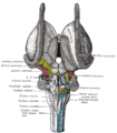
 Hind- and mid-brains; postero-lateral view. (Pulvinar visible near top.) | |
 Thalamic nuclei: MNG = Midline nuclear group AN = Anterior nuclear group MD = Medial dorsal nucleus VNG = Ventral nuclear group VA = Ventral anterior nucleus VL = Ventral lateral nucleus VPL = Ventral posterolateral nucleus VPM = Ventral posteromedial nucleus LNG = Lateral nuclear group PUL = Pulvinar MTh = Metathalamus LG = Lateral geniculate nucleus MG = Medial geniculate nucleus | |
| Details | |
| Part of | thalamus |
| Identifiers | |
| Latin | nuclei pulvinares (the nuclei plurally); pulvinar thalami (the set of nuclei singularly) |
| MeSH | D020649 |
| NeuroNames | 328 |
| NeuroLex ID | birnlex_824 |
| TA98 | A14.1.08.104 A14.1.08.610 |
| TA2 | 5665, 5698 |
| FMA | 62178 |
| Anatomical terms of neuroanatomy | |
The pulvinar nuclei or nuclei of the pulvinar (nuclei pulvinares) are the nuclei (cell bodies of neurons) located in the thalamus (a part of the vertebrate brain).[1] As a group they make up the collection called the pulvinar of the thalamus (pulvinar thalami), usually just called the pulvinar.
The pulvinar is usually grouped as one of the lateral thalamic nuclei in rodents and carnivores, and stands as an independent complex in primates.
Structure[edit]
By convention, the pulvinar is divided into four nuclei:
| TA alphanumeric identifier | TA name | English translation |
|---|---|---|
| A14.1.08.611 | nucleus pulvinaris anterior | anterior pulvinar nucleus |
| A14.1.08.612 | nucleus pulvinaris inferior | inferior pulvinar nucleus |
| A14.1.08.613 | nucleus pulvinaris lateralis | lateral pulvinar nucleus |
| A14.1.08.614 | nucleus pulvinaris medialis | medial pulvinar nucleus |
Their connectomic details are as follows:
- The lateral and inferior pulvinar nuclei have widespread connections with early visual cortical areas.
- The dorsal part of the lateral pulvinar nucleus predominantly has connections with posterior parietal cortex and the dorsal stream cortical areas.
- The medial pulvinar nucleus has widespread connections with cingulate, posterior parietal, premotor and prefrontal cortical areas.[2]
- The pulvinar also has input from the superior colliculus to inferior, lateral and medial sections, which seems to be important in the initiation and compensation of saccade,[3][4] as well as the regulation of visual attention[5][6]
Clinical significance[edit]
Lesions of the pulvinar can result in neglect syndromes and attentional deficits.[7] In addition, lesions in early life can impact normal visuomotor behaviors such as reaching and grasping.[8] Furthermore, the pulvinar was demonstrated to be instrumental in the preservation of vision afforded to a boy who lost his primary visual cortex bilaterally at birth[9] as well as other forms of blindsight in monkeys[10][11] and humans.[12] Strokes affecting the pulvinar have also been implicated in the development of chronic pain.[13] In a case study of photophobia caused by blue light, pulvinar nuclei associated with the melanopsin containing ipRGCs visual pathway where bilaterally activated.[14]
Other animals[edit]
The pulvinar varies in importance in different animals: it is virtually nonexistent in the rat, and grouped as the lateral posterior-pulvinar complex with the lateral posterior thalamic nucleus due to its small size in cats. In humans it makes up roughly 40% of the thalamus making it the largest of its nuclei.[15] Significant research has been undertaken in the marmoset examining the role of the retinorecipient region of the inferior pulvinar (medial subdivision), which projects to visual cortical area MT, in the early development of MT and the dorsal stream, as well as following early-life lesions of the primary visual cortex (V1).[16][17][18]
Etymology[edit]
The word pulvinar (/pʌlˈvaɪnər/) in Latin broadly means an armchair lined with numerous pillows. It was first neuroanatomically named by Karl Friedrich Burdach in 1817:[19] "The cushion (pulvinar), a swelling at the posterior end of the inner edge of the upper quadrigemina like a pillow over seats", English translation[20] (original German: "Das Polster (pulvinar), eine Anschwellung am hintern Ende des inner Randes der obern Vierhügel wie ein Kissen herüber legt"[19]). In Latin pulvinus could refer to "a sofa, cushioned seat, seat of honor, easy couch; of the couch or marriage-bed ", or more specifically, "a couch made of cushions, and spread over with a splendid covering, for the gods and persons who received divine honors; a couch or cushioned seat of the gods"[21] In the religion of ancient Rome, a pulvinar was an hetoimasia or empty throne, cushioned for occupation by a deity.[22] While anatomically, neuroanatomically there was no Roman deity between its arms, there was the pineal gland, that had in the 17th century, been identified by the French philosopher René Descartes as the seat of intellect and soul, and it has been suggested this link contributed to the first naming of this part of the brain by Karl Friedrich Burdach.[20]
References[edit]
- ^ Baud, RH; et al., "Latin index of TA98, Terminologia Anatomica version 1998", Federative International Programme on Anatomical Terminologies (FIPAT), International Federation of Associations of Anatomists (IFAA), hosted by the University of Fribourg (Switzerland)
- ^ Cappe, Céline; Morel, Anne; Barone, Pascal; Rouiller, Eric M. (September 2009). "The Thalamocortical Projection Systems in Primate: An Anatomical Support for Multisensory and Sensorimotor Interplay". Cerebral Cortex. 19 (9): 2025–2037. doi:10.1093/cercor/bhn228. PMC 2722423. PMID 19150924.
- ^ Berman, R. A.; Wurtz, R. H. (12 January 2011). "Signals Conveyed in the Pulvinar Pathway from Superior Colliculus to Cortical Area MT". Journal of Neuroscience. 31 (2): 373–384. doi:10.1523/JNEUROSCI.4738-10.2011. PMC 6623455. PMID 21228149.
- ^ Robinson, David Lee; Petersen, Steven E. (July 1985). "Responses of pulvinar neurons to real and self-induced stimulus movement". Brain Research. 338 (2): 392–394. doi:10.1016/0006-8993(85)90176-3. PMID 4027606. S2CID 7547426.
- ^ Petersen, Steven E.; Robinson, David Lee; Morris, J.David (January 1987). "Contributions of the pulvinar to visual spatial attention". Neuropsychologia. 25 (1): 97–105. doi:10.1016/0028-3932(87)90046-7. PMID 3574654. S2CID 23143322.
- ^ Chalupa, L. (1991). Visual function of the pulvinar. The Neural Basis of Visual Function. CRC Press, Boca Raton, Florida, pp. 140-159.
- ^ Arend, I.; Rafal, R.; Ward, R. (10 January 2008). "Spatial and temporal deficits are regionally dissociable in patients with pulvinar lesions". Brain. 131 (8): 2140–2152. doi:10.1093/brain/awn135. PMID 18669494.
- ^ Mundinano, Inaki (2018). "Transient visual pathway critical for normal development of primate grasping behavior". Proceedings of the National Academy of Sciences. 115 (6): 1364–1369. doi:10.1073/pnas.1717016115. PMC 5819431. PMID 29298912.
- ^ Mundinano, IC; Chen, J; de Souza, M; Sarossy, MG; Joanisse, MF; Goodale, MA; Bourne, JA (2017). "More than blindsight: Case report of a child with extraordinary visual capacity following perinatal bilateral occipital lobe injury". Neuropsychologia. 128: 178–186. doi:10.1016/j.neuropsychologia.2017.11.017. PMID 29146465. S2CID 207242249.
- ^ Kinoshita, Masaharu; Kato, Rikako; Isa, Kaoru; Kobayashi, Kenta; Kobayashi, Kazuto; Onoe, Hirotaka; Isa, Tadashi (December 2019). "Dissecting the circuit for blindsight to reveal the critical role of pulvinar and superior colliculus". Nature Communications. 10 (1): 135. Bibcode:2019NatCo..10..135K. doi:10.1038/s41467-018-08058-0. PMC 6329824. PMID 30635570.
- ^ Takakuwa, Norihiro; Isa, Kaoru; Onoe, Hirotaka; Takahashi, Jun; Isa, Tadashi (24 February 2021). "Contribution of the Pulvinar and Lateral Geniculate Nucleus to the Control of Visually Guided Saccades in Blindsight Monkeys". The Journal of Neuroscience. 41 (8): 1755–1768. doi:10.1523/JNEUROSCI.2293-20.2020. PMC 8115889. PMID 33443074.
- ^ Kletenik, Isaiah; Ferguson, Michael A.; Bateman, James R.; Cohen, Alexander L.; Lin, Christopher; Tetreault, Aaron; Pelak, Victoria S.; Anderson, Clark Alan; Prasad, Sashank; Darby, Richard Ryan; Fox, Michael D. (February 2022). "Network Localization of Unconscious Visual Perception in Blindsight". Annals of Neurology. 91 (2): 217–224. doi:10.1002/ana.26292. PMC 10013845. PMID 34961965. S2CID 245553461.
- ^ Vartiainen, Nuutti; Perchet, Caroline; Magnin, Michel; Creac’h, Christelle; Convers, Philippe; Nighoghossian, Norbert; Mauguière, François; Peyron, Roland; Garcia-Larrea, Luis (March 2016). "Thalamic pain: anatomical and physiological indices of prediction". Brain. 139 (3): 708–722. doi:10.1093/brain/awv389. PMID 26912644.
- ^ Panorgias, Athanasios; Lee, Danielle; Silva, Katie E.; Borsook, David; Moulton, Eric A. (2019). "Blue light activates pulvinar nuclei in longstanding idiopathic photophobia: A case report". NeuroImage: Clinical. 24: 102096. doi:10.1016/j.nicl.2019.102096. PMC 6879998. PMID 31795037.
- ^ LaBerge, D. (1999). Attention pp. 44-98. In Cognitive science (Handbook of Perception and Cognition, Second Edition), Bly BM, Rumelhart DE. (edits). Academic Press ISBN 978-0-12-601730-4 p. 73
- ^ Warner, Claire E.; Kwan, William C.; Bourne, James A. (28 November 2012). "The Early Maturation of Visual Cortical Area MT is Dependent on Input from the Retinorecipient Medial Portion of the Inferior Pulvinar". Journal of Neuroscience. 32 (48): 17073–17085. doi:10.1523/JNEUROSCI.3269-12.2012. PMC 6621860. PMID 23197701.
- ^ Warner, Claire; Goldshmit, Yona; Bourne, James (2010). "Retinal afferents synapse with relay cells targeting the middle temporal area in the pulvinar and lateral geniculate nuclei". Frontiers in Neuroanatomy. 4: 8. doi:10.3389/neuro.05.008.2010. PMC 2826187. PMID 20179789.
- ^ Warner, Claire E.; Kwan, William C.; Wright, David; Johnston, Leigh A.; Egan, Gary F.; Bourne, James A. (16 February 2015). "Preservation of Vision by the Pulvinar following Early-Life Primary Visual Cortex Lesions". Current Biology. 25 (4): 424–434. doi:10.1016/j.cub.2014.12.028. PMID 25601551.
- ^ a b Burdach, K.F. (1817). Ueber die Aufgabe der Morphologie (in German). In der Dyk'schen Buchhandlung. Retrieved 2023-08-29.
- ^ a b Turliuc, Dana; Turliuc, Şerban; Cucu, Andrei; Dumitrescu, Gabriela Florenţa; Cărăuleanu, Alexandru; Buzdugă, Cătălin; Tamaş, Camelia; Sava, Anca; Costea, Claudia Florida (2016). "A review of analogies between some neuroanatomical terms and roman household objects". Annals of Anatomy. 204: 127–133. doi:10.1016/j.aanat.2015.07.001. ISSN 0940-9602.
- ^ "Charlton T. Lewis, Charles Short, A Latin Dictionary, pulvīnar". Perseus Digital Library. Retrieved 2023-08-29.
- ^ Vranesevic, Branka; Spehar, Olga (2021). "Enthronement of the invisible. Understanding the origin and evolution of the iconography of empty thrones and hetoimasia in the late antique period". Zograf (45): 1–14. doi:10.2298/zog2145001v. ISSN 0350-1361.
Additional images[edit]
-
 Thalamus
 -
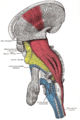 Deep dissection of brain-stem. Lateral view.
 -
 Dissection of brain-stem. Dorsal view.
 -
 Scheme showing central connections of the optic nerves and optic tracts.
 -
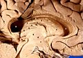 Human brain left dissected midsagittal view


External links[edit]
- Stained brain slice images which include the "pulvinar" at the BrainMaps project
- Atlas image: eye_38 at the University of Michigan Health System - "The Visual Pathway from Below"